BIOLOGÍA - GEOLOGÍA . COM

1st ESO

3rd ESO

4th ESO

Biology 2nd Baccalaureate

Index by courses

Forums


Skip navigation

4.4.1.2. Secondary structure of proteins

Secondary structure of proteins

The secondary structure of a protein is the arrangement in space of the amino acids sequence or primary structure. It is due to the rotational capacity of the bonds that form the α-carbon, and to the radicals of the amino acids, which make the polypeptide acquire a stable spatial arrangement, the secondary structure.

The secondary structure of the polypeptide chain depends on the amino acids that make it up.

It is the highest structural level that fibrous proteins reach.

Three types of secondary structure are known:

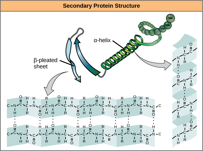
α-helix

The secondary structure in the α-helix is formed by helically winding the peptide chain on itself, clockwise (clockwise).

This type of structure is typical of proteins that have a high number of amino acids with large or hydrophilic radicals, since the charges interact with the water molecules that surround it. The structure is stabilized due to the large number of links by hydrogen bonds established between the -NH- of an amino acid and the -C=O of the fourth amino acid that follows the spiral. The α-helix has 3.6 amino acids per turn.

For this reason, in the α-helix, the oxygens of all the –C=O groups are oriented in the same direction, and the hydrogens of all the -NH groups are oriented in just the opposite direction, while the radicals of the amino acids they are directed towards the outside of the α-helix.

The side chains of amino acids do not intervene in the bonds, so proteins with very different primary structures can have the same secondary structure.

Some amino acid side chains can react with others with a charge of the same sign, and destabilize the structure, so that areas in which the α-helix is ​​not appreciated can be found in the same protein.

Alpha-helix.jpg
By Alejandro Porto - Derivada de File:AlphaHelixProtein.jpg de Maksim e imagen generada de archivo pdb con Jmol (un visor Java de código abierto para estructuras químicas en tres dimensiones. http://www.jmol.org/) ., CC BY-SA 3.0, Link

Conformation β or folded sheet

Another way of arranging the primary structure of a protein in space is the β conformation or folded sheet, such as that found in β-keratin, present in nails, hair and feathers.

It is a zig-zag- shaped structure, forced by the rigidity of the peptide bond and the apolarity of the radicals of the amino acids that make up the molecule. Several chains are arranged in parallel, forming a folded sheet.

It stabilizes by creating hydrogen bonds between the –C=O and –NH groups of the parallel chains. There are no hydrogen bonds between the nearby amino acids of the polypeptide chain.

Figure 03 04 07

By CNX OpenStax [CC BY 4.0], via Wikimedia Commons

Collagen helix

In addition to the α-helix and the folded sheet, there are other types of secondary structures, such as the triple helix of collagen.

It is the secondary structure that collagen presents, which is part of the tendons and connective tissues; it is a particularly rigid structure.

Collagen has a special helix arrangement, somewhat more elongated than the α-helix, due to the abundance of glycineproline, and hydroxyproline. These amino acids have a structure that makes the formation of hydrogen bonds very difficult, which is why an α-helix is ​​not formed, but rather a left-handed helix somewhat more stretched than the α-helix, having only three amino acids per turn.

Collagen is formed by the association of three left-handed helices, which wind between them in a clockwise direction, and are joined by hydrogen brigde type bonds.

By Vossman. Modificado por Alejandro Porto (File:Collagentriplehelix.png) [CC BY-SA 3.0], via Wikimedia Commons

Fundamental ideas about the secondary structure of proteins

The Secondary structure of a protein is the arrangement in space of the peptide chain.

  • α helix: the amino acid chain is wound in a clockwise turn. Hydrogen bonds form between the O of the –C=O of an amino acid and the H of NH of the fourth amino acid that follows. The α-helix has 3.6 amino acids per turn.

In the case of the collagen helix, it is wound in a left-handed way and there are three amino acids per turn. 

  • Beta conformation: A helix is ​​not formed, but the peptide chain adopts a zigzag shape due to the rigidity of the peptide bond, without the presence of hydrogen bonds between amino acids of the same chain. If several chains are arranged in parallel, hydrogen bonds can appear between amino acids, forming a folded sheet


Legal warning

Blog

Download

Contact


Follow us if it has been useful to you biologia-geologia.com.
 

Biology and Geology teaching materials for Compulsory Secondary Education (ESO) and Baccalaureate students.