BIOLOGÍA - GEOLOGÍA . COM

1st ESO

3rd ESO

4th ESO

Biology 2nd Baccalaureate

Index by courses

Forums


Skip navigation

9.1.4. The eye and vision

The sense of sight

The sense of sight is probably the most important of those we have, since it gives us most of the information we receive. It allows us to recognize the color, shape, size and distance of an object.

If you blindfold yourself, you will realize how difficult it is to understand what is going on around you. Blind people have had to develop other senses, such as hearing or touch, to be able to adapt to their environment.

The eye is an exteroceptor sensory organ (receives information from outside) and a photoreceptor (receives light stimuli). This information is transmitted to the brain where it is processed and produced a response.

The receptors of the eye are in a very complex organ, the eye, comprising the eyeball and some accessory organs, such as muscle, which give high mobility, and glands that lubricate and protect.

Partes del ojo

Eye anatomy

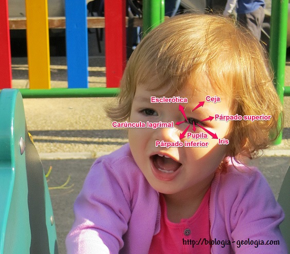
The eyeball is very fragile, so it is protected by a cavity in the skull called the orbit, and by a series of accessory organs such as:

  • The eyebrows, hairs that cover the prominences located above the eyes, which deflect sweat preventing it from entering the eyes .
  • The eyelids are folds of the skin that cover the front of the eye, distribute tears and prevent the entry of foreign objects and drying out.
  • The eyelashes are hairs located on the edge of the eyelid. They filter the light making it more diffuse .
  • The lacrimal glands are structures that secrete tears that prevent the eye from drying out. Tears contain lysozyme, which is a substance with bactericidal properties.

The eye muscles allow you to move the eye in different directions to be able to direct your eyes towards any object you want to look at .

Partes del ojo

By Gabrielzerrisuela (Own work) [CC BY-SA 3.0], via Wikimedia Commons

The eye is made up of different parts , each with a specific function:

  • Conjunctiva: membrane that covers and protects the front of the eyeball and the inside of the eyelids.
  • Sclera: hard, white outer layer that protects the eyeball and gives it shape and consistency. The front part is transparent, and is called the cornea.
  • Choroid: located below the sclera. It is dark in color, highly irrigated to nourish the rest of the structures.
  • The iris, behind the cornea, is a muscle that presents various colors (brown, blue, green, ...) and regulates the passage of light through a central hole, the pupil.
  • Ciliary muscle: attached to the choroid and the lens, it is ring-shaped and serves to curve the lens to focus on objects.
  • Crystalline: small, transparent and elastic structure, shaped like a biconvex lens that allows objects to be focused on the retina. The lens separates two spaces occupied by:
    • Aqueous humor: liquid that occupies the space between the cornea and the lens.
    • Vitreous humor: gelatinous substance that occupies the space between the retina and the lens, also transparent, maintains the shape of the eyeball and a uniform surface of the retina so that the reception of images is clear.
  • Retina : layer of cells that lines the inside back of the eyeball. It captures light stimuli and transmits the information to the optic nerve . It is made up of two types of photoreceptor cells:
    • Cones: they are responsible for daytime vision, visual acuity and color vision. There are three types of cones: those that are sensitive to red light, blue light and green light.
    • Rods: they do not need a lot of light intensity to get excited, but they do not differentiate colors. They allow us to have night vision, being able to see something at night. Surely you have noticed that at night we only see shadows, in black and white, without being able to distinguish the colors.

Other parts of the retina of special interest are:

  • Fovea: area of ​​the retina where cones are grouped, so visibility is maximum. It is located in front of the center of the lens, the pupil, and the cornea, and is where images of objects are reflected.
  • Blind spot: area of ​​the retina where the optic nerve exits, so there are no photoreceptor cells and it does not receive any visual information.
  • Optic nerve: formed by extensions of nerve cells that transmit information from photoreceptor cells to the brain.

3D model: Anatomy of the eye.

Activity: Parts of the eye.

Activity: The eye.

Activity: The eye.

Activity: Dissection of a cow's eye.

Game: Parts of the eye.

Functioning of the eye

The light that penetrates the interior of the eyeball passes through the cornea, and the lens focuses the image on the retina, where the photoreceptor cells (rods and cones) are excited and transmit the nerve impulse, through the optic nerves , to the brain.

The image that is formed on the retina is an inverted image (upside down) and smaller than reality, although the brain interprets this information and makes us see it in its real position. Remember that the sense organs only transform the different stimuli into nerve impulses, and it is the nervous system that interprets the information received. It is in the visual cortex that the sensation of seeing occurs.

Light intensity regulation

The pupil, by contracting or relaxing some muscles of the iris, is responsible for regulating the amount of light that reaches the retina.

In low light, the iris relaxes and the pupil increases in size to let in as much light as possible.

If there is a lot of light, the iris contracts to prevent too much light from reaching the retina and can damage the photoreceptor cells.

Image focus

The lens accommodates itself, curving more or less according to the distance to which objects are located so that the image is focused correctly (although inverted) on the retina. Thus, if the object is far away, the ciliary muscle causes the lens to flatten, and if it is close, to bulge.

Interactive activity: Accommodation of the eye.

Stereoscopic vision

The stereoscopic vision is the ability to integrate into a single three - dimensional image, emboss the two images we receive from each of our eyes, located one on the other side. Although the eyes are very close together, we see the object from different angles. This information is transmitted to the brain, where the visualized image is processed and created.
Eye diseases

The eye is a very sensitive organ whose function can be altered for many reasons. Some of the main visual diseases are:

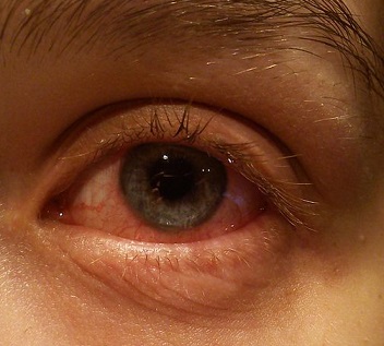
  • The conjunctivitis is an inflammation of the conjunctiva of the eye caused by infections or allergies. It causes irritation, itching, pain and stinging.

Conjuntivitis

By Daemonanyndel (Own work) [CC BY-SA 3.0], via Wikimedia Commons

  • The cataracts occur when the lens becomes opaque and the light does not let pass. It begins with blurred vision decreasing vision, but can end in blindness.

Cataratas: cuando el cristalino se vuelve opaco

By Rakesh Ahuja, MD (Own work) [GFDL, CC-BY-SA-3.0 or CC BY-SA 2.5-2.0-1.0], via Wikimedia Commons

  • The myopia causes distant objects appear blurred. This occurs because the eyeball is longer than normal and the images are formed in front of the retina. The myopia is the inability to focus on objects far because the lens is too convex and can not stretch to focus. It is corrected with divergent lenses.
  • The hyperopia occurs when the eyeball is shorter than normal, then the image of near objects is formed behind the retina. It is the inability to focus on nearby objects because, contrary to what happens in myopia, the lens is too stretched and cannot bulge. It is corrected with converging lenses.
  • The astigmatism is due to a deformation in the curvature of the cornea, causing objects look distorted. Vertical lines look distorted because the lens bulges unevenly. It is corrected using cylindrical lenses.

  • The presbyopia or eyestrain: visual acuity loss. It prevents you from seeing nearby objects because the lens hardens and loses the ability to focus.
  • The color blindness. All the diseases of the eye that we have seen were related to problems in the lens, but in this case it is the cones, which make it difficult to distinguish colors such as red and green.

Answer in your notebook

9.5.- What is the difference between myopia and hyperopia?

Answer in your notebook

9.6.- What is the function of the lens?

Answer in your notebook

9.7.- With which receivers are we able to see during the day? And at night?


Legal warning

Blog

Download

Contact


Follow us if it has been useful to you biologia-geologia.com.
 

Biology and Geology teaching materials for Compulsory Secondary Education (ESO) and Baccalaureate students.